Stay informed with the latest surgical insights, clinical updates, technological advancements, and expert guides from Siora Surgicals.
A Spinal Fixation System is a group of implants that helps stabilize the vertebral column during healing. It may include screws, rods, plates, cages, or connectors. These components are placed near damaged or unstable spinal segments. They can limit painful movement while bone fusion develops.
In the operating room, a surgeon first identifies the affected levels through imaging and direct assessment. Screws anchor into selected vertebrae. Rods then connect those anchors and support the intended alignment. In some procedures, a cage restores disc height and creates space for bone growth. The system does not “repair” every spinal problem by itself. It supports a carefully planned biological process.
As spine surgeon Dr. Edward C. Benzel has stated, “The spine is a dynamic structure, not a static one.” That idea explains why fixation requires balance. Too little support may allow instability. Too much rigidity may increase stress on nearby segments. The best construct depends on bone quality, anatomy, symptoms, and surgical goals.
Details matter.
A patient’s posture can change the plan. So can osteoporosis, previous surgery, or a narrow spinal canal. Modern systems offer useful precision, but they are not risk-free. Infection, nerve injury, implant loosening, and adjacent-segment problems remain possible. The evidence is strong for selected cases, yet not every back-pain patient needs fixation. This distinction is sometimes overlooked. A reliable explanation must acknowledge uncertainty, because successful treatment involves more than inserting hardware.
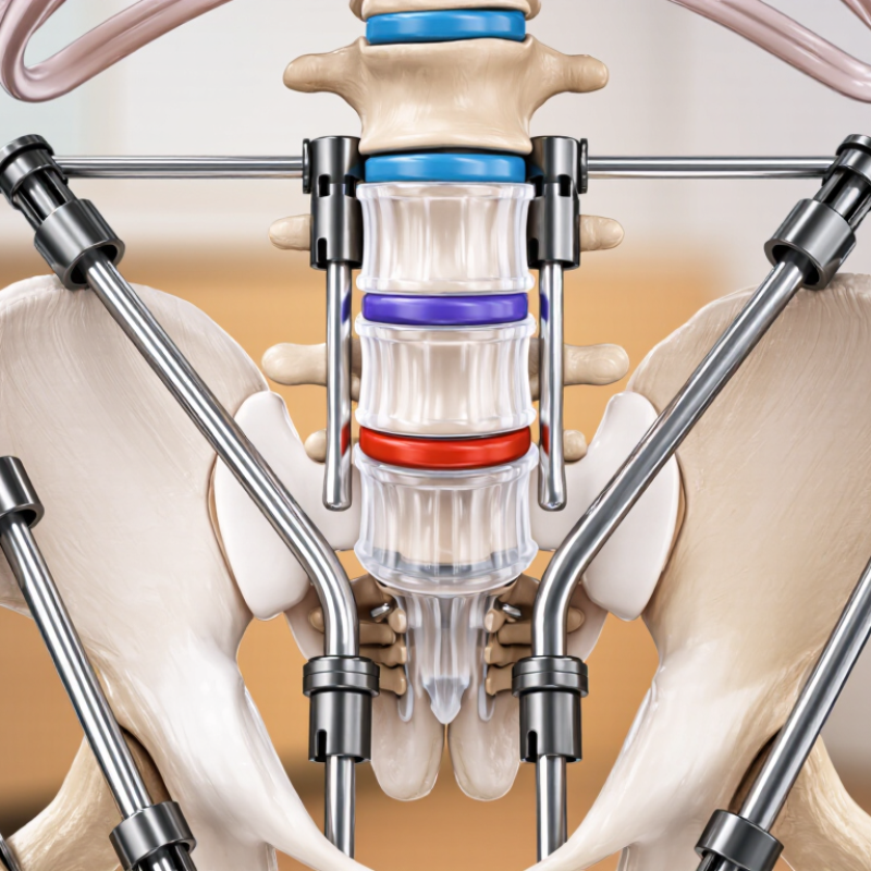
A spinal fixation system is a group of implants that stabilizes vertebrae during healing. It commonly includes screws, rods, plates, connectors, and sometimes cages. Surgeons place these components around an unstable spinal segment. The structure limits painful movement while bone graft gradually forms a stronger fusion. Think of it as an internal support frame, not a replacement spine.
Fixation may be considered after fractures, deformity, infection, tumors, or severe instability. The decision depends on imaging, symptoms, bone quality, medical history, and surgical goals.
However, widespread back pain does not automatically require fixation. The biology is not perfectly predictable. Fusion can fail, and nearby spinal levels may later experience greater stress. Professional assessment remains essential.
Ask what problem the system is designed to solve. Request a clear explanation of each implant and its material. Discuss bone health, smoking, rehabilitation, and warning signs. The American Academy of Orthopaedic Surgeons notes that recovery varies by procedure and patient factors. Follow-up imaging helps assess alignment and bone growth. A second opinion can also be reasonable when the treatment plan feels unclear.
A spinal fixation system supports vertebrae that cannot remain stable on their own. It commonly includes screws, rods, plates, or connecting devices. Surgeons place these components around the affected spinal segment. The hardware limits abnormal movement while bone healing or fusion develops. The goal is stability.
Why is spinal fixation used? It may be recommended after a spinal fracture, severe deformity, or significant vertebral instability. It can also support the spine after decompression surgery, when removing bone or disc material creates extra movement. In some degenerative conditions, fixation helps maintain alignment and protects a fusion area. A patient may notice less painful motion when standing, turning, or walking.
Pain alone is insufficient. A careful decision considers neurological symptoms, imaging findings, spinal balance, bone quality, and overall health. Magnetic resonance imaging can show nerve pressure, while X-rays may reveal movement between vertebrae. Computed tomography often clarifies fractures and bone structure. Surgeons also compare non-surgical treatments and the risks of hardware failure, infection, nerve injury, or nonunion. The decision is rarely perfect. Symptoms and scans do not always match neatly. Good care requires honest discussion, realistic recovery expectations, and follow-up examinations to confirm that the spine is healing as intended.
A spinal fixation system uses mechanical parts to stabilize a weakened or surgically treated section of the spine. Its design depends on the patient’s anatomy, the affected levels, and the surgical goal. Not every system uses every component.
Pedicle screws are commonly placed into the vertebrae through carefully planned paths. They provide strong anchor points. Long rods connect these screws and help maintain alignment while bone healing develops. Set screws secure the rods in place. Small connectors may join rods, adjust their position, or support more complex spinal shapes. In some procedures, plates and screws are used near the front of the spine instead.
Interbody cages are placed between vertebral bodies after a damaged disc is removed. They help restore disc height and create space for bone growth. Surgeons may add bone graft material inside or around the cage. The graft supports fusion, while the fixation hardware limits unwanted movement. It is a quiet partnership.
In real clinical practice, the system must fit more than an X-ray. Bone quality, nerve location, posture, and movement all matter. A technically sound plan can still require adjustment during surgery. Labels and component names may also vary between manufacturers, which can create confusion. Careful imaging, sterile technique, and the surgeon’s experience guide selection and placement. Nakne
This chart shows an illustrative bilateral one-level posterior fixation construct. Component counts vary according to the spinal level, surgical technique, and patient anatomy.
How it works: Pedicle screws anchor the construct to the vertebrae. Longitudinal rods connect the screws and help maintain alignment. Set screws secure the rods to the anchors, while a transverse connector may improve rotational stability when included.
A spinal fixation system stabilizes vertebrae that are weak, unstable, or healing after surgery. It usually includes screws, rods, plates, or connecting devices. The surgeon selects each component according to the patient’s anatomy, diagnosis, and bone quality. Placement is not a one-size-fits-all procedure.
The operation begins with anesthesia and careful positioning on a padded surgical table. The surgeon makes an incision over the affected spinal levels and gently moves muscles aside. Imaging helps identify the correct vertebrae before any hardware is inserted. Small screws are then placed through specific parts of the vertebrae. Their position may be checked with real-time X-ray or computer-assisted navigation. This matters near the spinal cord and nerve roots.
Rods or plates connect the screws and create a stable framework. The surgeon may also remove pressure from nerves or add bone graft material to support fusion. Intraoperative monitoring can help detect changes in nerve function, although it cannot remove every risk. No operation is perfectly predictable. Bone density, anatomy, scar tissue, and movement can complicate placement. Afterward, imaging confirms alignment, while the clinical team checks strength, sensation, wound condition, and pain. Recovery differs widely, and early symptoms should be reported rather than dismissed.
A spinal fixation system supports healing by limiting unwanted movement between unstable vertebrae. It usually combines screws, rods, plates, or interbody cages. Surgeons place these components to restore alignment and share mechanical loads. The goal is not to make the spine immovable forever. It is to protect the surgical area while bone grows across the intended fusion site.
The biology is less tidy. Bone still decides. Fixation can reduce painful micromotion, maintain disc height, and give bone graft a stable environment. The North American Spine Society’s clinical guidance emphasizes patient selection, imaging, and regular follow-up because hardware cannot correct every cause of back pain. A 2019 Agency for Healthcare Research and Quality HCUP report ranked spinal fusion among the most common inpatient operating-room procedures in the United States. That volume reflects clinical need, not guaranteed success.
Healing may take months, and smoking, diabetes, poor bone density, or excessive early loading can interfere with fusion. The World Health Organization reported that low back pain affected about 619 million people globally in 2020, showing why careful treatment decisions matter.
Stability must also preserve useful movement above and below the construct. This balance is easy to overlook. Surgeons therefore assess symptoms, neurological findings, bone quality, and postoperative imaging rather than relying on hardware alone. The evidence is strong in many situations, but not perfect; some patients continue to experience pain despite solid fusion.
It is an internal support frame for an unstable spinal segment. It may include screws, rods, plates, connectors, and cages. The system is not a replacement spine. It limits painful movement while bone healing develops.
Fixation may support the spine after a fracture, deformity, infection, tumor, or severe instability. It may also help maintain alignment after decompression surgery. Pain alone is not enough reason. Symptoms and scans must be considered together.
The implants reduce unwanted movement between unstable vertebrae. They can maintain alignment and support a bone graft. This creates a steadier environment for fusion. Bone still decides the outcome.
No. The goal is not permanent rigidity. The system protects the healing area while bone grows across the fusion site. Movement above and below the construct remains important. This balance can be easy to overlook.
X-rays can show spinal alignment and movement between vertebrae. Magnetic resonance imaging may reveal pressure on nerves. Computed tomography can clarify fractures and bone structure. Doctors also review symptoms, bone quality, and medical history.
Healing may take several months. Recovery depends on the procedure, bone health, general health, and rehabilitation. Follow-up imaging can check alignment and bone growth. Progress is not always smooth.
Smoking, diabetes, poor bone density, and excessive early loading may slow fusion. Patients should discuss bone health and rehabilitation before surgery. Following activity instructions matters. Healing can still be uncertain.
Possible concerns include hardware failure, infection, nerve injury, and nonunion. Some people continue to feel pain despite solid fusion. Nearby spinal levels may later experience greater stress. The scans and symptoms may not match neatly.
Ask what problem the system is intended to solve. Request a clear explanation of every implant and its material. Discuss expected recovery, warning signs, rehabilitation, and follow-up imaging. A second opinion may help when the plan feels unclear.
A Spinal Fixation System is a medical device assembly used to stabilize and align sections of the spine when bones, discs, or supporting structures are damaged or unstable. It may be recommended for conditions such as fractures, deformities, spinal degeneration, or instability that cannot be adequately managed through non-surgical treatment. By holding the affected vertebrae in a controlled position, the system helps reduce unwanted movement, relieve mechanical stress, and support the natural healing process.
The system commonly includes screws, rods, plates, connectors, and sometimes bone graft material, depending on the surgical plan and the patient’s condition. During surgery, a qualified medical team places the fixation components into or around selected vertebrae using imaging and careful anatomical guidance. The hardware maintains spinal alignment while the bone gradually heals and fuses when fusion is intended. Although fixation can improve stability and function, the appropriate method, risks, and expected recovery vary for each patient and should be discussed with a spine specialist.