Choosing among Tarsal Fixation Nails is not simply a brand contest. The right implant depends on the bones involved, the fracture or fusion pattern, and the patient’s anatomy. A nail that looks suitable on a product page may not match the space available in a small tarsal bone.
This guide examines what makes a fixation nail worth considering. It looks at dimensions, material, screw placement, insertion tools, and compatibility with imaging. These details matter in practice: a few millimeters can affect fit, while screw direction can influence how securely bone fragments are held. Surgeons also consider bone quality, soft-tissue condition, and whether the implant supports the planned procedure. There is no universal winner.
“Top” needs context. A design used for one hindfoot procedure may not suit another, and availability or clinical evidence can differ by region. Product descriptions are useful, but they cannot replace peer-reviewed studies, manufacturer instructions, or a surgeon’s assessment. This comparison therefore focuses on features and selection questions rather than claiming one nail is best for everyone. That distinction is easy to overlook.
Readers will find practical criteria for comparing options, along with limitations to keep in mind. Evidence for specific designs may be uneven, and published results do not always predict an individual outcome. The details deserve a closer look. A qualified foot-and-ankle specialist can explain which implant fits a specific diagnosis, anatomy, and surgical plan.
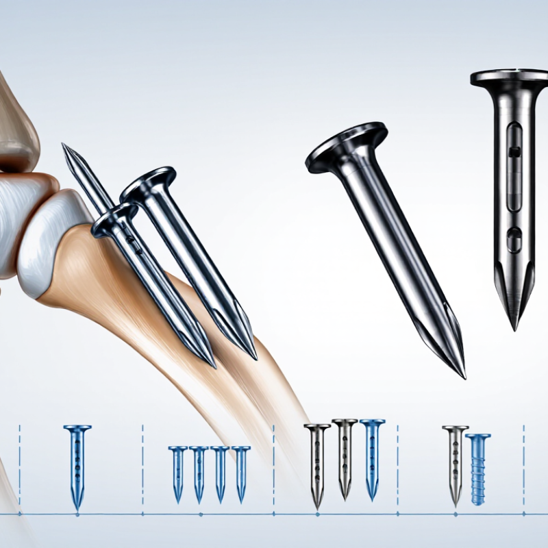
What Tarsal Fixation Nails Are and How They Stabilize the Foot
Tarsal fixation nails are metal implants used to hold selected foot and ankle bones steady. A common hindfoot nail passes through the heel bone and into the lower leg. Locking screws help control rotation and limit movement while bones heal or fuse. The goal is a stable, well-aligned foot, not simply a rigid one.
Surgeons may consider a nail for complex fractures, severe deformity, or ankle and hindfoot fusion. The exact design and path depend on the bones involved, bone quality, and the patient’s needs. Imaging helps guide placement and check alignment. A nail can provide strong internal support, but it cannot guarantee healing. Swelling, infection, poor bone healing, or pressure from the implant may still occur. No device fits every foot; that point can be easy to overlook when comparing options.
Tips: Ask which joints the nail will cross, how long weight-bearing may be restricted, and what follow-up images will check. Keep the questions specific. Recovery varies, and a planned schedule may need adjustment if pain or healing changes.
What Are the Top Tarsal Fixation Nails? - What Tarsal Fixation Nails Are and How They Stabilize the Foot
| Nail Design | Typical Construct | How It Stabilizes the Foot | Common Uses | Key Considerations |
|---|---|---|---|---|
| Retrograde tibiotalocalcaneal (TTC) nail | A nail is inserted from the plantar heel and passes through the calcaneus and talus into the tibia. Locking screws secure the construct in the bones. | Spans the ankle and subtalar joints, limiting motion while fusion develops and helping resist bending and rotational forces. | Selected cases of severe ankle and hindfoot arthritis, deformity, or instability when fusion of both joints is planned. | Requires suitable bone, a carefully planned entry point, and assessment of alignment. It sacrifices motion at the joints being fused. |
| Compression-capable TTC nail | A TTC nail with a mechanism designed to apply compression across one or both fusion sites; the mechanism differs by implant design. | Compression brings prepared bone surfaces together, while the nail and locking screws maintain alignment and provide internal support. | Selected TTC arthrodesis procedures where the surgeon considers controlled compression appropriate. | Compression capability does not guarantee union. Bone preparation, fixation, alignment, and patient factors remain important. |
| Short hindfoot nail | A retrograde nail that crosses the hindfoot fusion area and ends in the distal tibia. | Provides an internal load-sharing support across the hindfoot while locking screws help control rotation and translation. | Selected hindfoot or TTC fusion cases where a shorter tibial segment is suitable. | Available fixation points and the amount of tibial purchase vary by design. Bone quality and the planned fusion level guide selection. |
| Long hindfoot nail | A retrograde nail that extends farther up the tibial canal than a short nail. | Engages a longer segment of the tibia and can provide a longer internal lever arm for construct support. | Selected complex reconstructions or cases in which the surgeon wants fixation extending farther proximally. | Requires evaluation of the tibial canal, alignment, and any existing implants or deformity. Greater length is not automatically preferable. |
| Static locking configuration | A hindfoot nail secured with locking screws in fixed positions, without an active compression step during insertion. | Locks the bones relative to the nail to help maintain the intended position during healing. | Fusion procedures where stable positioning is the main fixation objective. | Screw number, direction, and placement depend on the nail system and anatomy. Stability also depends on bone quality and surgical technique. |
Note: “Tarsal fixation nail” commonly refers to a retrograde hindfoot intramedullary nail, especially a TTC nail. There is no universally ranked “top” design; implant choice depends on the diagnosis, anatomy, bone quality, planned fusion, and the treating surgeon’s assessment. This table is for general education and is not a substitute for medical advice.
FAQS
It is a metal implant that steadies selected foot and ankle bones. A common hindfoot nail passes through the heel bone toward the lower leg.
It may be considered for complex fractures, severe deformity, or ankle and hindfoot fusion. The plan depends on the bones involved and the patient’s needs.
Locking screws help limit rotation and shifting. Some designs also compress prepared bone surfaces. Stable alignment is the goal, not stiffness for its own sake.
No. Longer nails may link the tibia, talus, and calcaneus. Shorter constructs can target fewer joints. The planned fusion guides the choice.
Nail length, diameter, curvature, and screw angles matter. The heel entry point may irritate nearby soft tissue if it is prominent. Small details matter.
No single design suits every anatomy or surgical goal. Bone quality and imaging findings also affect selection. Standard options can fit awkwardly.
No. It provides internal support, but healing is not guaranteed. Swelling, infection, poor bone healing, or implant pressure may still occur.
Ask which joints the nail will cross, how long weight-bearing may be restricted, and which follow-up images are planned. Keep questions specific. Plans may change. Pain or healing changes can affect recovery.
Conclusion
Tarsal Fixation Nails are internal devices used to stabilize bones in the hindfoot or midfoot when a fracture, deformity, or joint condition requires support. Placed through the foot, a nail can help hold bones in alignment and encourage healing or fusion. Designs vary in length, diameter, locking options, and screw configuration, allowing fixation to be adapted to the anatomy and the treatment goal. The choice of nail depends on imaging, bone quality, the location and severity of the problem, overall health, and the surgeon’s assessment.
During surgery, the surgeon positions the nail and secures it with screws to maintain stability. Potential benefits include improved alignment and support, but risks may include infection, nerve or blood-vessel injury, delayed healing, hardware irritation, or the need for further treatment. Recovery varies: patients may need a period of limited or no weight-bearing, followed by gradual rehabilitation and follow-up imaging. The expected outcome depends on the underlying condition, healing progress, and adherence to the care plan.